


Orlando hospital uses 3D-printed models of fetuses to prep for in-utero surgery
New modeling technology allows surgeons to visualize complicated predelivery surgery to correct birth defects
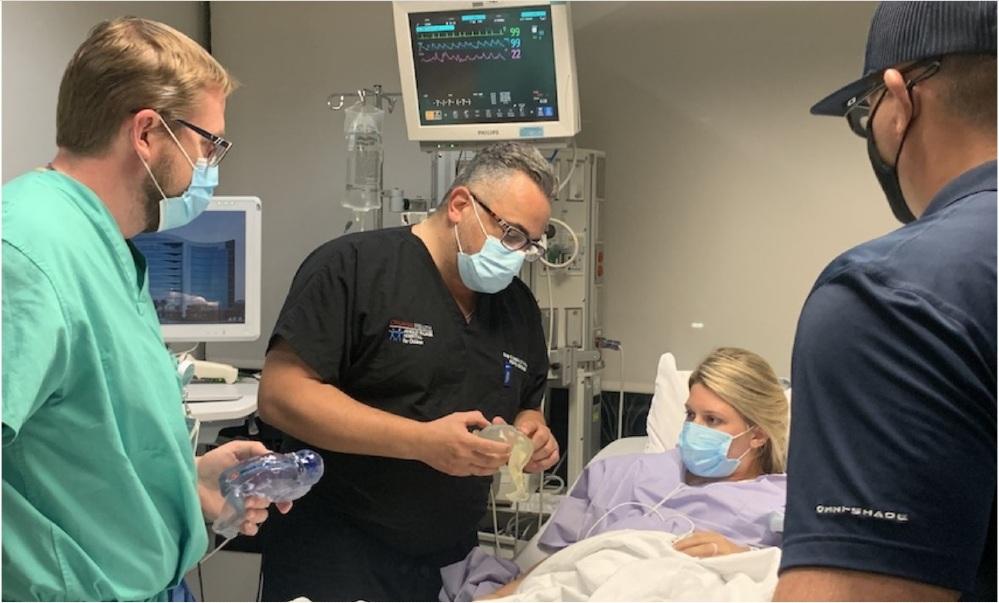
Orlando Health pediatric neurosurgeon Dr. Samer Elbabaa shows expectant parents Jocelyn and Jared Rodriguez (right) a 3D-printed model of their developing baby. Orlando Health surgeons use models to prepare for in-utero procedures to correct spina bifida, a birth defect that occurs when the spinal cord fails to close normally during development. Images: Orlando Health
The following report was prepared by Orlando Health.
A state-of-the-art in-utero procedure allows surgeons to correct birth defects of developing babies still in the womb. But operating on a mother and her unborn child at the same time can be challenging and unpredictable.
To provide its world-class surgeons additional information prior to surgery, Orlando Health Winnie Palmer Hospital for Women & Babies in Florida uses MRI, ultrasound imaging, and 3D printing technology to create a first-of-its-kind, detailed model of the fetus. The model allows surgeons to plan procedures ahead of time and helps them identify potential obstacles and reduce risks that can arise during surgery.
Models are currently being used to plan for in-utero surgery that repairs spina bifida, a birth defect that occurs when the spinal cord fails to close normally during development. The condition can cause a lifetime of neurological disabilities, including an inability to walk.
“The 3D reconstruction of the fetus can really educate the surgeon on the real-life shape, size, and location of the spinal lesion, as well as prepare the surgeon to have the appropriate equipment ready to treat this condition surgically,” said Samer Elbabaa, M.D. Elbabaa, the medical director of pediatric neurosurgery at Orlando Health, added, “It’s a level of detail that we are not able to see in traditional imaging, but that is extremely valuable in these cases where we cannot actually see the defect ahead of surgery.”
To create the models, Orlando Health works with the prototyping and 3D printing team at Digital Anatomy Simulations for Healthcare LLC (DASH), the Orlando, Fla., company that developed the technology.
Crude, single-color items have been 3D-printed in the past, but DASH has taken the process to the next level, developing technology to enhance MRI and ultrasound images taken throughout the pregnancy to reconstruct accurate curves and edges. DASH prints the high-resolution models with multiple colors and materials, allowing surgeons to see details such as skeletal structure, nerve and vascular anatomy, and fluid sacs in the spine and brain caused by spina bifida.
The models are currently being used in the hospital’s fetal surgery program, which has performed 25 procedures since it began in 2018. Orlando Health is one of only 12 facilities in the U.S. and the only one in Florida able to perform this kind of surgery.
“The fetal models not only help surgeons plan for things like where to make an incision and how to repair the defect, but also help reduce the duration of the surgery to limit the developing baby’s exposure,” said DASH President and CEO Jack Stubbs. “We are able to create models that are extremely realistic by using a stack of two-dimensional images taken throughout the pregnancy and enhancing them to reconstruct a better visualization of what the fetus truly looks like.”
The 3D-printed models give surgeons a clearer picture of what to expect during surgery and also allow them to better explain the baby’s condition to parents and show what the child’s treatment will entail.
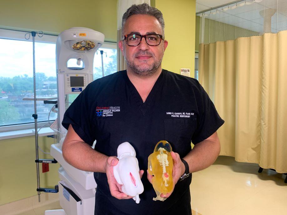
Dr. Elbabaa holds 3D-printed models of fetuses. They let surgeons review, visualize, and prepare for a complex procedure normally supported only by MRI and ultrasound imaging.
For first-time parents Jared and Jocelyn Rodriguez, the model made them more confident about moving forward with their daughter’s surgery.
“At first we just thought it was a model showing the same kind of condition that our baby was diagnosed with, but then Dr. Elbabaa told us that it was made using the 20-week MRI of our daughter,” Jared Rodriguez said. “We could see the brain and the spine and I looked down at it and thought, ‘I’m holding my daughter right now? That’s pretty awesome.’”
The parents say although they are prepared for the challenges their daughter may face, they’re glad this technological development is helping to give her a healthier future. “Every appointment we go to, we just keep getting more good news and she’s already showing how strong she is,” Jocelyn Rodriguez said. “We know that this surgery will give her the best shot at a normal lifestyle, and we’re excited to see the positive results as she grows.”
Surgeons are seeing successful results from fetal surgery for spina bifida. Most babies who undergo the procedure experience significantly fewer health issues and greater functionality than those who receive surgery after they’re born. Some of the first patients to undergo the procedure are now learning to walk on their own.
Experts hope to expand the program in the future by modeling other types of birth defects that can be treated during fetal surgery.
Click here for a video report on the procedure and parents Jared and Jocelyn Rodriguez.
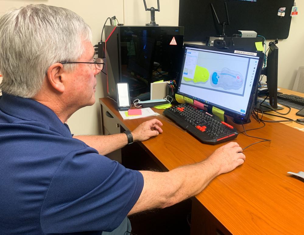
Digital Anatomy Simulations for Healthcare President and CEO Jack Stubbs prepares a 3D model of a fetus using MRI and ultrasound imaging. The printed model will give Orlando Health surgeons a life-size representation they can use to prepare for in-utero surgery.
About the Publication
- Podcasting

In this episode of The Fabricator Podcast, Caleb Chamberlain, co-founder and CEO of OSH Cut, discusses his company’s...
- Trending Articles